
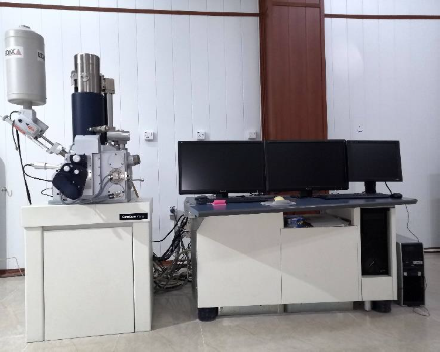
Scanning Electron Microscope Laboratory

In this laboratory we use scanning electron microscope to image samples of different materials and microorganisms by scanning them with a focused beam of electrons.
Firstly, the specimens were put in the sputter coater to coat by a thin layer of gold for more reflection of electrons. Secondly, the sample put in the SEM chamber to image in different directions, and it shape, size, structure and features are appeared with different magnifications until 250 times than that of light microscope.
Hemin Muhammad
Phone: +964 770 1574136
Email: [email protected]